Treatment Strategy for ecDNA-Driven Tumors Shows Potential
, by Carmen Phillips
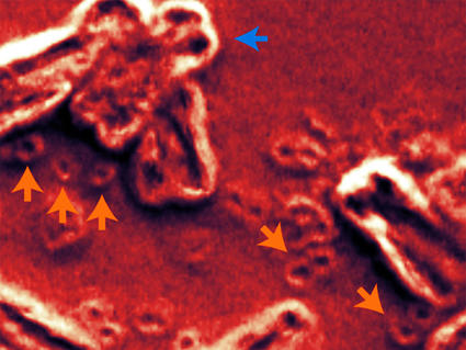
Researchers have identified a way to potentially kill tumors fueled by extrachromosomal DNAs (ecDNAs), free-floating circular chunks of DNA suspected of making cancers more aggressive and helping them thwart all manner of treatments.
With the aid of an experimental drug, scientists have now shown that they can take advantage of an instability in cancer cells created by ecDNAs.
Findings from the study were published November 6 in Nature, along with two others on ecDNAs funded by the Cancer Grand Challenges, an international initiative sponsored by NCI and Cancer Research UK.
One of the other studies shows that ecDNAs are found in many more cancers and at much higher levels than previously thought, while the third provides new details about how ecDNAs are passed on in cancer cells as tumors grow. (See below, “More Insights on ecDNAs.”)
“We’ve learned that ecDNAs are a huge problem in cancer,” said Paul Mischel, M.D., of Stanford University, the leader of the Cancer Grand Challenges ecDNA team (called eDyNAmiC) and a senior investigator on all three studies.
In the treatment-focused study, Dr. Mischel and his colleagues showed that ecDNAs can also cause problems in cancer cells, in the form of serious difficulties copying their DNA in preparation to divide into two new cells. And an experimental drug, called BBI-2779, could exploit those difficulties, they found. In mice with tumors carrying ecDNAs with a cancer-causing genetic mutation, combining BBI-2779 with a drug that targets the mutation quickly shrank tumors and kept them at bay for relatively long periods.
In the study, each drug alone modestly reduced tumor size, Dr. Mischel explained. But, he continued, “when you put the two [drugs] together you get a massive response.”
A closely related drug—called BBI-355—is already being tested in humans in a small clinical trial, both alone and in combination with one of several targeted therapies, depending on the genetic changes that are present in participants’ tumors.
What’s known about ecDNAs in cancer
Although ecDNAs were first discovered more than 60 years ago, until recently they were a biological mystery. In part, that’s because finding them and analyzing their makeup was very difficult.
However, with the aid of numerous advanced technologies, a more complete portrait of ecDNAs is emerging.
For example, it’s been well established that only cancer cells have ecDNAs and that they appear to influence cancer cells in important and diverse ways, explained Ian Fingerman, Ph.D., of NCI’s Division of Cancer Biology.
“The more we dig into ecDNAs, the more important they seem to be,” Dr. Fingerman said.
He pointed, for example, to research led by Dr. Mischel and his Stanford colleague Howard Chang, M.D., Ph.D., which showed that ecDNAs almost always contain cancer-causing genes, known as oncogenes. They also often house many copies of the same oncogene, known as gene amplification, and those genes tend to be very active.
This hyperactivity is partly due to the circular structure of ecDNAs, explained Bishoy Faltas, M.D., of Weill Cornell Medicine, who led a separate NCI-funded study of ecDNAs in bladder cancer published in the same issue of Nature. “The circular shape allows the oncogenes on ecDNA to interact more readily with other elements on the ecDNA, leading to increased activity of those genes,” Dr. Faltas said.
A few years ago, Drs. Chang, Mischel, and their team made a related and surprising discovery. Much like teenage boys at their first dance, they found, ecDNAs tend to cluster in large groups, or what they call “hubs.” The individual ecDNAs in each hub are physically tethered together, with their close proximity creating a “cooperative environment” in which components on one ecDNA molecule can, and often do, control the activity of components on another.
By hosting amplified oncogenes, Dr. Faltas noted, ecDNAs are well suited to helping cancer cells do things like quickly adapt to and fend off treatments. In his team's recent study, for example, they showed that oncogenes on ecDNAs appeared to be “the keys to [bladder tumors'] resistance to chemotherapy,” he explained.
The finding is in-line with others that strongly suggest ecDNAs appear to provide cancer cells and the tumors of which they are apart with some significant advantages. But Dr. Mischel and his team wondered whether the oncogene hyperactivity in ecDNAs might also have some disadvantages for tumors, including something that could be a potentially exploitable weakness.
Taking advantage of conflict
For the study involving BBI-2779, Dr. Mischel and his team started with a hunch. Because of the high levels of oncogene amplification in ecDNAs, they suspected that cells with ecDNAs should also have high rates of events called transcription-replication conflicts.
These conflicts are literal physical collisions among molecules in the nucleus involved in the processes of transcription, during which an RNA copy of a segment of DNA is made, and DNA replication, which is the copying of DNA in preparation for cell division.
Both transcription and replication are in high gear in cancer cells, so they suspected that there should be plenty of these conflicts, as well as an associated condition called replication stress.
And when replication stress gets severe enough, it can lead to DNA that is so badly damaged it causes cells to die.
They tested their hunch by analyzing cancer cells with ecDNAs that had amplification of a well-known oncogene called MYC and amplification of the same gene on its appropriate chromosome.
They found precisely what they expected to: very high levels of transcription of MYC on ecDNAs compared with its chromosome-bound counterpart, an abundance of evidence of transcription-replication conflicts and replication stress, and plenty of DNA damage.
Exploiting replication stress by blocking CHK1
In the face of such tumultuous conditions, normal cells typically die. But in the case of cancer cells, which are particularly good at skirting death, the researchers suspected that these cells might need an added push.
That push was blocking a protein called CHK1, which acts like a traffic cop in the cell nucleus, slowing or stopping cell division when DNA damage is detected.
That’s where BBI-2779 comes in: It blocks CHK1 from performing its duties. In lab experiments, treating ecDNA-containing cancer cells with BBI-2779 caused the equivalent of multiple multi-car pileups, jamming up the transcription and replication machinery and causing mass cell death.
In mice, the drug on its own modestly shrank stomach tumors of ecDNA-containing cancer cells with mutations in an oncogene called FGFR. Treating the mice with infigratinib, which targets cells with FGFR mutations, shrank tumors for a short time, but they quickly regrew as the ecDNA-containing cells ramped up FGFR activity faster than infigratinib could shut it down.
Combining BBI-2779 with infigratinib looked to be the winning recipe, thwarting this adaptation and keeping tumors from regrowing.
One Trial Underway, Much More Research to Do
The clinical trial of BBI-355 is funded by Boundless Bio, which Drs. Mischel and Chang founded. Both now serve on its scientific advisory board.
The trial is the first toe in the water of human studies of ecDNA-directed treatments, Dr. Mischel noted. There are likely to be other ways of targeting ecDNA-fueled tumors, he continued.
According to Dr. Faltas, there may also be opportunities to prevent ecDNA from forming in the first place. In his group’s study, for example, the appearance of ecDNA was linked to the development of mutations caused by an enzyme called APOBEC3. This enzyme can also cause powerful breaks in DNA, he explained, such as those that can fragment it and potentially lead to the formation of structures like ecDNA.
This finding “suggests that APOBEC3-induced mutations either precede or occur at the same time” as ecDNAs are being formed, Dr. Faltas said. “So, you start to think, can we intercept the formation of ecDNAs?”
Dr. Mischel and his Cancer Grand Challenges colleagues are thinking along the same lines, he explained, with studies already focused on identifying the early events in ecDNA formation and developing methods for monitoring ecDNAs in blood samples.

